The PhenoImager Fusion microscope can function as a standalone ultrafast slide scanner ideal for standard throughput and high-plex spatial phenotyping applications. It has fluorescence and brightfield capabilities and can separate up to 7 colors with a resolution up to 0.25 um/pixel.
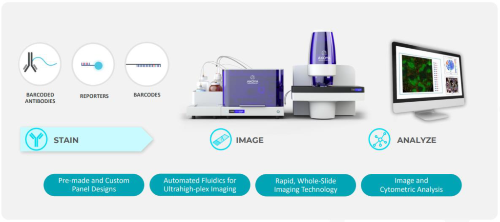
- A comprehensive end-to-end solution containing the fluidics platform, assay reagents, and bioinformatics
- Scalable and capable of imaging 6 biomarkers at high throughput to more than 50+ biomarkers per run
- Isolates autofluorescence from tissue samples for more accurate and quantifiable imaging
- Preserves the sample for downstream Region Of Interest (ROI) analysis and H&E staining
Tissue Types
- Mouse Fresh Frozen
- Human Fresh Frozen
- Human FFPE
- TMAs (tumor microarrays)
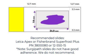
Sample Preparation and workflow:
Sections can be placed on standard, positively charged histology slides. Tissues must fit within the imageable area below.
PhenoCycler Staining Workflow (50+ antibodies):
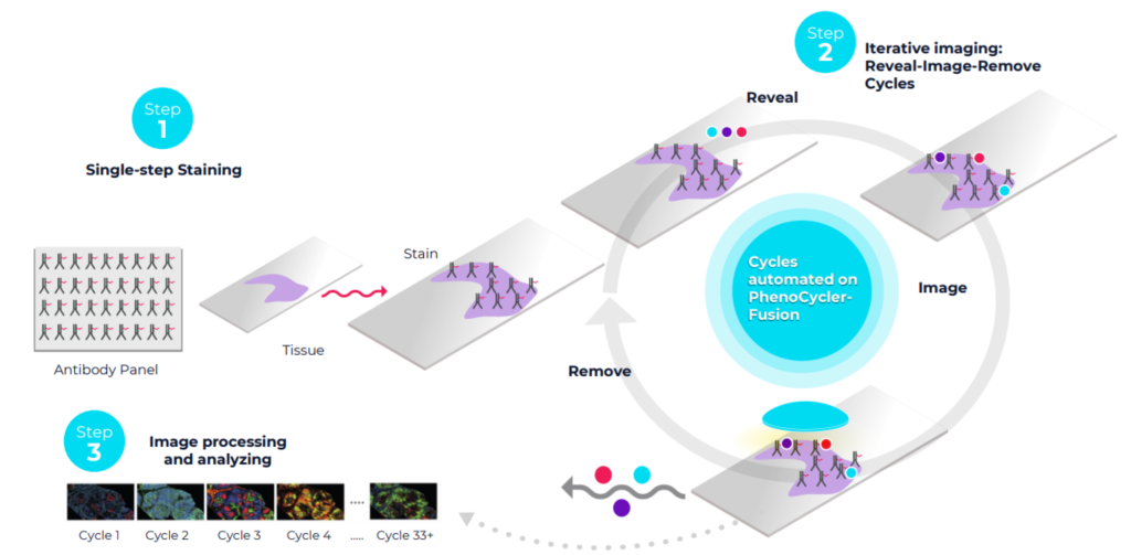
PhenoCycler staining uses antibodies attached to a unique oligonucleotide barcode. All antibodies are stained in a single cocktail, and the automated system then adds 3 complementary reporters at a time with a fluorophore which specifically detects each unique antibody. The signals are revealed, imaged, and gently removed, and the process repeats until all the markers are imaged.
Akoya Resources
Technical resources: Tissue processing best practices, Tissue Staining procedure, molecular barcoding workflow, etc – https://www.akoyabio.com/support/reagents/
PCF Screened Antibody Database: https://www.akoyabio.com/phenocycler/assays/antibodies/
Explore interactive datasets: https://www.akoyabio.com/fusion/data-gallery/
Using the PhenoCycler-Fusion 2.0:
The first step is to contact the facility at huixu@illinois.edu and briefly describe the project. Next, we will schedule a meeting to discuss your needs, clarify the sample type, number, and antibody panel needed, and ask you to submit an order request on FBS.